
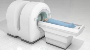
Positron emission tomography, commonly known as PET scanning, is a nuclear medicine imaging technique that produces three-dimensional images of functional processes in the body. Positron Emission Tomography Scanners uses radioactive tracers, called radiotracers or radiopharmaceuticals, to produce images of organs and tissues as they function.
How does PET Scanning Work?
PET scanning works by introducing tiny amounts of radiotracers into the body, usually through intravenous injection. These radiotracers involve substances that mimic normal body chemicals and are attached to a short-lived radioactive atom, often fluorine-18 or carbon-11. As the radiotracer travels through the part of the body being examined, it gives off positrons (positive electrons). The positrons interact with electrons, producing two 511-keV gamma rays emitted at 180° to each other. Circular arrays of detectors surrounding the patient’s body capture these gamma rays. A computer analyzes the information from the detectors and produces a 3D color image of the tracer concentration in the body. Areas with a higher concentration of radiotracer appear brighter on the scan, indicating more activity in that area. The images can identify areas where cells are more or less active than normal.
Common Radiotracers Used in PET Scanning
Fluorodeoxyglucose (FDG): Fluorodeoxyglucose, marked with fluorine-18, is the most common radiotracer used in PET scans today. FDG is taken up by cells with a high glucose metabolism, such as cancer cells and active immune cells. It is useful for imaging various cancers, cardiovascular disease, and certain infections and inflammatory conditions.
Fluorodopamine (FDA): Fluorodopamine, marked with fluorine-18, is used as a PET radiotracer for diagnosing Parkinson’s disease and other movement disorders. It binds to dopamine receptors in the brain, helping evaluate loss of dopaminergic neurons.
Fluoromisonidazole (FMISO): Fluoromisonidazole, marked with fluorine-18, is used for assessing oxygen levels in tissues. Since hypoxic (oxygen-starved) cells take up more FMISO than normoxic cells, it can help detect hypoxic regions in tumors. This aids in diagnosis and treatment planning for cancers.
Acetate: Acetate, labeled with carbon-11, is taken up by the prostate and can help detect prostate cancer recurrence after initial treatment. It is also useful for imaging cardiac function.
Ammonia: Ammonia, labeled with nitrogen-13 or fluorine-18, aids evaluation of blood flow to the heart muscles. It binds reversibly to myocardium in direct proportion to regional blood flow. Myocardial perfusion imaging with ammonia PET can help diagnose coronary artery disease.
Types of PET Scans and their Clinical Applications
Whole body PET scan: A whole body PET scan captures images from the skull to the mid-thighs. It is commonly used to stage and monitor many cancers like lung, breast, colorectal, melanoma, and lymphoma.
Brain PET scan: Brain PET scanning uses specialized scanners that focus on the brain region. It helps diagnose and monitor brain diseases like Alzheimer’s, epilepsy, stroke, brain tumors, and traumatic brain injury. Brain PET with FDG and florbetapir scans are FDA-approved for evaluating Alzheimer’s disease.
Cardiac PET scan: Also known as myocardial perfusion imaging, cardiac PET scanning evaluates blood flow to the heart muscles using radiotracers like rubidium-82 and ammonia. It is useful for diagnosing coronary artery disease and guiding revascularization procedures like angioplasty.
PET/CT fusion imaging: Many centers now offer integrated PET/CT scanners, which perform both PET and low-dose CT scans simultaneously. The CT scan provides morphological (anatomical) detail to correctly localize functional abnormalities seen on PET. PET/CT is especially useful for cancers that have spread, helping pinpoint the primary and metastatic tumors. It also reduces scanning time compared to standalone PET and CT scans.
Advantages of PET Scanning
Some key advantages of PET imaging include:
– Functional imaging: PET scans provide information about physiological processes at the molecular and cellular levels, unlike CT/MRI which only show anatomical details. This makes PET well-suited for cancer, neurological disorders, and cardiac evaluation.
– Sensitivity: PET scans can detect metabolic changes in very small structures and at early disease stages when anatomical changes may not yet be visible on other scans. This improves detection rates.
– Quantification: The uptake and distribution of radiotracers can be objectively measured on PET scans, allowing quantitative analysis of biological processes. This aids diagnosis, staging, treatment monitoring, and clinical research.
– Non-invasive: PET scanning does not involve any needles or incisions and uses very low radiation doses comparable to other nuclear medicine procedures. It causes minimal discomfort during the scan.
– Co-registration with CT/MRI: Integrated PET/CT scanners help accurately localize areas of abnormal tracer uptake seen on PET, resolving any ambiguity around anatomical localization of functional lesions.
*Note:
1. Source: Coherent Market Insights, Public sources, Desk research
2. We have leveraged AI tools to mine information and compile it