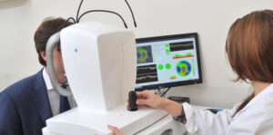
Optical coherence tomography (OCT) is a medical imaging technique that uses light waves to capture micrometer-resolution, cross-sectional images from within biological tissues. In just over 20 years, Optical Coherence Tomography Devices has progressed from a novel concept to a routinely used clinical tool and is considered a breakthrough technology for non-invasive evaluation of the eye, skin and cardiovascular system. This article aims to provide an overview of OCT technology, its applications and recent advances.
How does OCT Work?
OCT captures micrometre-scale resolution, cross-sectional images from within optical scattering media (e.g., biological tissue) using low-coherence interferometry. It detects back-scattered light from within tissues and constructs 2D or 3D images. A near-infrared light source is used with a coherence length of typically 5-15 micrometres, which allows reflections to be discriminated from different depths. By acquiring multiple A-scans (i.e. depth profiles), a cross-sectional tomogram can be constructed or multiple adjacent tomograms combined to produce a 3D volumetric dataset. Like ultrasound imaging or MRI, OCT provides depth imaging capability but with a higher resolution and without the need for ionizing radiation.
Clinical Applications of OCT in Ophthalmology
OCT revolutionized ophthalmology and is now the standard of care for retinal pathology diagnosis and glaucoma management. It allows non-invasive high-resolution visualization of retinal layers and can detect subtle changes not visible on examination. Cross-sectional retinal images provide crucial anatomical information to diagnose a wide range of retinal pathologies like macular edema, epiretinal membranes, retinal detachments and tumors. OCT-based thickness maps of the retinal nerve fiber layer aid in glaucoma diagnosis and monitoring. Recent advances like enhanced depth imaging OCT detect choroidal features enabling new insights into diseases like age-related macular degeneration. OCT angiography visualizes the retinal and choroidal microvasculature without contrast injection.
Cardiovascular and Non-Ocular Applications
Intravascular OCT (IVOCT) delivers high resolution cross-sectional images of coronary arteries and can identify thin-cap fibroatheromas, which are vulnerable plaque morphologies associated with acute coronary syndromes. It aids interventions by providing additional anatomical detail beyond angiography alone. Other non-ophthalmic applications include nasal cavity imaging for sinus tumors/polyps, otoscopy of middle ear structures and laryngotracheal examination for airway diseases. Emerging areas include imaging of esophageal diseases and gastrointestinal cancers using endoscopic OCT. Its micrometer resolution and absence of ionizing radiation make OCT an attractive tool for longitudinal monitoring of treatment response in oncology.
Technical Advances in OCT Systems
Recent technical innovations have expanded OCT capabilities. Swept source OCT uses rapidly tunable laser sources instead of broadband light, enabling faster imaging speeds up to 400,000 A-scans/sec. This allows larger volumes to be imaged in shorter times and permits dynamic in-vivo imaging. Full-field OCT captures a width of tissue simultaneously for high throughput. Optical frequency domain imaging (OFDI) uses frequency-swept interferometry, delivering imaging speeds rivalling swept source OCT. Functional extensions integrate Doppler OCT for microvasculature flow measurements, polarization sensitive OCT for collagen/tissue structure evaluation and spectroscopic OCT for chemical/molecular analysis. Miniaturized probes now enable endomicroscopic and ophthalmic applications. Most recent developments include multi-beam and adaptive optics OCT to overcome scattering/aberrations and achieve even higher resolutions.
Advanced Applications of OCT
Advanced OCT applications are pushing the technology into new territories. Compact OCT systems mounted on surgical microscopes or endoscopes provide real-time intraoperative guidance. OCT enabled optical biopsy facilitates minimally-invasive tissue characterization. Structural and molecular OCT signatures may aid disease diagnosis from tissue micro-architecture alone. Optical metrology utilizes OCT’s precision to map nanoscale surfaces and micro-geometry. Functional extensions permit dynamic in-vivo assessment of nanomechanical properties in tissues/cells. Emerging developments encompass cell/organ culture growth studies, microfluidic chip analysis, micro-electromechanical systems testing as well as pre-clinical research spanning regenerative medicine to pharmaceutical development. With ongoing technical progress, OCT’s potential is expanding to excite new discoveries in science and applications in healthcare.
Conclusion
Since its invention over 25 years ago, Optical Coherence Tomography Devices has emerged from obscurity to become a mainstream clinical imaging modality across ophthalmology as well as certain vascular and non-vascular specialties. It delivers unparalleled, micrometer-scale resolutions non-invasively by pure optical means. Initial applications in ophthalmology revolutionized management of vitreo-retinal disorders. However, the wider scope of OCT continues to unfold with technological innovations that enhance imaging speeds and functional capabilities. Its various clinical applications are expected to proliferate by integration into endoscopes, catheters and surgical microscopes, further cementing OCT as an indispensable tool for medicine in the coming years. With ongoing technical progress, OCT remains a highly active area of research promising new discoveries and breakthrough clinical applications.
*Note:
1. Source: Coherent Market Insights, Public sources, Desk research
2. We have leveraged AI tools to mine information and compile it
