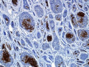
Immunohistochemistry (IHC) is a technique that utilizes the principles of antibodies and antigen-antibody binding reactions to detect antigens in cells of a tissue section. It allows for the visualization of antigens via linked detection systems and microscopes and offers the ability to examine the distribution and level of target antigens in different types of cells and tissues. IHC relies on antibodies produced in animals that are specific for antigens of interest. These primary antibodies can identify a wide range of molecular targets, such as proteins, sugars, DNA, and nucleic acids that exist within tissues.
Sample Preparation and Antibody Incubation
The first key steps in IHC involve preparing the sample tissue, usually from surgical biopsies or autopsy material, and fixing it to stabilize the cellular components so they retain antigenicity for analysis. Common fixatives include neutral buffered formalin and alcohols. Next, the tissue sample undergoes processing, embedding in paraffin wax, and sectioning into very thin slices that are then placed onto glass slides for IHC staining. After deparaffinization and rehydration, the tissue sections are treated to recover or unmask antigens by heat-induced epitope retrieval or protease enzymes. Next, endogenous peroxidases that could cause non-specific background staining are blocked. The tissue sections are then incubated with the chosen primary antibody with specificity for the desired antigen target.
Signal Amplification and Visualization
After primary antibody binding, sections are washed to remove any unbound primary antibody. Then a secondary antibody linked to an enzyme or other detectable molecule is applied. This secondary antibody is specific for the species that produced the primary antibody, such as anti-mouse or anti-rabbit. A signal amplification step may be included using a labeled polymer conjugated to multiple enzyme molecules for increased sensitivity. Finally, the signal is developed using a chromogen substrate that produces a visible colored precipitate where the target antigen exists. Sections are counterstained with hematoxylin to show nuclei of cells for orientation. The slides can then be observed using light microscopy to determine the cell types and localization of the target antigen.
Applications of Immunohistochemistry
IHC has a myriad of applications in research, diagnostics, and therapeutics. Some key uses include:
Disease Diagnosis – IHC can help diagnose cancers and other diseases by identifying cell types and markers. Example diagnostic markers include estrogen/progesterone receptors in breast cancer or keratins in epithelial tumors.
Prognostic Factors – Certain markers have prognostic value. For instance, HER2/neu status predicts response to HER2-targeted therapies for breast cancer patients. Ki-67 staining determines tumor proliferation rates.
Molecular Classification – IHC divides tumors into molecular subtypes based on marker expression, aiding treatment selection. For example, lung adenocarcinomas can have KRAS, EGFR, or ALK abnormalities.
Forensic Pathology – IHC plays an important role in determining cause of death, distinguishing tissue types, and aiding identification. For example, CD68 stains macrophages in post-mortem exams.
Research Applications – Techniques like multiplex-IHC and imaging mass cytometry expand IHC capabilities for cell phenotyping, functional assays, and protein interaction mapping in disease models and normal tissues.
Limitations and Considerations for Immunohistochemistry
While IHC is a highly valuable research and diagnostic tool, there are some limitations to consider:
Fixation Effects – Conditions like fixation duration impact antigenicity of targets. Standardization is important between cases.
Antibody Specificity – Cross-reactivity or low affinity antibodies may cause false-positives or lack of signal even with real antigens present.
Technical Variability – Staining protocols need optimization and normalization between technicians for consistency.
Antigen Loss – Some targets are more susceptible to loss during processing, reducing sensitivity.
Quantitation Challenges – Scoring relies on visual assessment and is more subjective than quantitative methods.
Interpretation Complexity – Heterogeneous expression patterns require experienced evaluation, especially with multiplexing approaches.
Despite these caveats, immunohistochemistry remains an indispensable technique in pathology and biomedical research due to its ability to map molecular features in situ within tissues and cells. With appropriate controls and validation, it provides powerful insights into health, disease, and therapeutic responses.
*Note:
1. Source: Coherent Market Insights, Public sources, Desk research
2. We have leveraged AI tools to mine information and compile it
