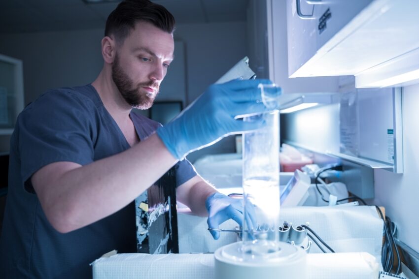
Nuclear medicine is a medical specialty that uses small amounts of radioactive material, known as radioisotopes or radiotracers, to diagnose and treat diseases. By visualizing physiological functions inside the body, diagnostic radioisotopes are revolutionizing healthcare and helping doctors gain a better understanding of various medical conditions. Let’s take a closer look at these valuable medical tools.
What are Diagnostic Radioisotopes?
Diagnostic Radioisotopes are unstable forms of elements that emit radiation as they undergo radioactive decay. Some commonly used radioisotopes for imaging include technetium-99m, fluorine-18, iodine-123, and gallium-67. These radioisotopes are attached to biologically active molecules known as radiopharmaceuticals or radiotracers. Once introduced into the body, the radiotracer travels through blood and accumulates in specific organs or tissues based on their metabolic or functional characteristics. By detecting the radiation signals emitted by the radioisotopes with specialized scanning devices, physicians can generate detailed images of internal organs and assess their structure and function.
Nuclear imaging modalities used include positron emission tomography (PET), single-photon emission computed tomography (SPECT), and gamma scanning. These techniques play a crucial role in medical diagnostics as they provide valuable insights into physiological and pathological abnormalities that may not be detectable by other means. Some diseases that are commonly evaluated using diagnostic nuclear medicine procedures include cancer, heart disease, infections, neurological disorders, and several others.
Applications in Oncology
Cancer diagnostics and management have greatly benefited from nuclear medicine technology. One of the most widely used radioisotopes in oncology is fluorine-18 (18F), which is incorporated into radioactive glucose analog radiotracers like fluorodeoxyglucose (18F-FDG). Tumor cells are known to utilize elevated levels of glucose for growth and proliferation. By detecting areas of increased 18F-FDG uptake on PET scans, physicians can screen for tumors, evaluate the extent of spread, detect recurrence after treatment, and monitor response to therapy.
Certain PET radiopharmaceuticals target tumor-specific molecular processes and receptors to detect certain cancers. For example, gallium-68 labelled prostate specific membrane antigen (68Ga-PSMA) PET is highly sensitive for detecting recurrent prostate cancer. 131Iodium is commonly used for imaging and treating differentiated thyroid cancers by taking advantage of iodine accumulation in thyroid cells. New radiotracers that home in on molecular targets like PSMA, somatostatin receptors, and fibroblast activation protein continue to expand the scope and accuracy of nuclear medicine in cancer care.
Cardiology and Neurology Applications
Diagnostic Radioisotopes techniques help examine heart anatomy, function, blood flow, and viability. SPECT imaging with radioisotopes like thallium-201 and technetium-99m is useful for detecting coronary artery disease by evaluating perfusion and wall motion abnormalities. PET radiotracers like rubidium-82 and ammonia-13N provide valuable physiological information about myocardial blood flow. These tests aid in diagnosing ischemic heart disease, assessing heart damage, and guiding cardiac interventions.
Several neurologic disorders can also be evaluated using radiotracers that bind to specific receptors and accumulate in brain regions. For example, 18F-FDG PET scans map glucose metabolism changes to detect conditions that alter brain activity patterns like epilepsy, Alzheimer’s disease, Parkinsonism, and brain tumors. Dopaminergic neurotransmission imaging with radiotracers like 18F-DOPA is helpful in diagnosing movement disorders. 123I-ioflupane (DATSCAN) SPECT aids in distinguishing Parkinson’s disease from other parkinsonian syndromes. These nuclear medicine procedures have transformed neurologic disease characterization and management.
Infection Scanning
Certain radiopharmaceuticals are helpful in diagnosing and localizing infections in the body. For example,technetium-99m labelled leukocytes or antibiotics concentrate in sites of infection after intravenous administration. SPECT/CT scans using such radiotracers allow diagnosis of uncertain focal infections like osteomyelitis, cardiac or vascular graft infections that may not be apparent on other studies. Gallium-67 citrate SPECT aids in evaluation of fever of unknown origin by localizing inflammatory foci. Such infection scans provide crucial clinical information to guide patient management decisions.
Advancing Diagnosis with Hybrid Imaging
The integration of nuclear imaging modalities with anatomical X-ray computed tomography (CT) is a major advance, allowing accurate localization of radiotracer uptake within the body. Hybrid imaging systems like SPECT/CT and PET/CT combine functional and anatomical data to facilitate precise diagnosis. Correlating radiotracer accumulation patterns on PET or SPECT images with underlying CT anatomy greatly enhances diagnostic confidence in detecting and characterizing diseases. With continual technological developments and new radiotracer development, diagnostic nuclear medicine is poised to play an increasingly important role in multidisciplinary healthcare by offering non-invasive insights into physiology at the molecular level.
In summary, radioisotopes and radiotracers have transformed medical diagnosis by enabling visualization of physiological processes in vivo. With further refinements, nuclear medicine imaging promises to refine disease characterization, guide treatment decisions, help develop individualized care plans, and monitor therapeutic responses—ultimately advancing patient care. Diagnostic radioisotopes exemplify the benefits that technology can deliver to improve human health and wellbeing when guided by scientific progress and medical insights.

